Highlights

Sunday, August 29, 2010

Cataract Zoo 4: Christmas Tree Cataract


A Christmas Tree Cataract is comprised of refractile needles located in the deep cortex of the lens. They crisscross the lens fibres reflecting light in different colours, looking very much like tinsel on a christmas tree..hence the name. 

The cause is thought it be from the age related breakdown of lens crystallins induced by elevated calcium levels. 

G. Adrien Shun-Shin* Gijs F. J. M. Vrensen,\ Nicholas P. Brown,* Ben Willekens,^
Marianne H. Smeets,^ and Anthony J. Bron*)

Cataract Zoo 3: Posterior Supcapsular

Posterior Subcapsular Cataract:
The greasy reflection off the back surface of the lens is the subcapsular opacity, and was visually devastating. This patient is diabetic and while this was the worst eye -there were similar changes in their other eye. This type of lens opacity is a common consequence of Diabetes. Modern surgical techniques gave this patient spectacular vision once again.

Cataract Zoo 2 : Dendritic Cataract

Dendritic Cataract:
This is a congenital cataract. An opacity in the sutures of the lens often has intricate branches. This one is beautifully tinged with azure. The vision is only one letter different in this eye. 

Cataract Zoo 1: Cortical

Cortical Changes:  Cataracts are ubiquitous - often the first obvious signs are cortical.

Cortical changes revealed by dilated pupil
Vesicles: fluid filled bubbles
More Bubbles: retro-illuminated
Vesicles with some sutural spokes (and a spec of posterior subcapsular change): 2 lines VA reduction


Myelinated Nerve Fibres

The insulating and protective fatty myelin sheath, which surrounds our nerves to protect and support them, doesn't generally enter the eye.
This myelination of the optic nerve develops embryologically in a posterior to anterior direction. The nerves are roughly myelinated in the intracranial and intracanal portions of the optic nerve by the seventh month (after conception) but doesn't usually reach the lamina cribosa until full term.
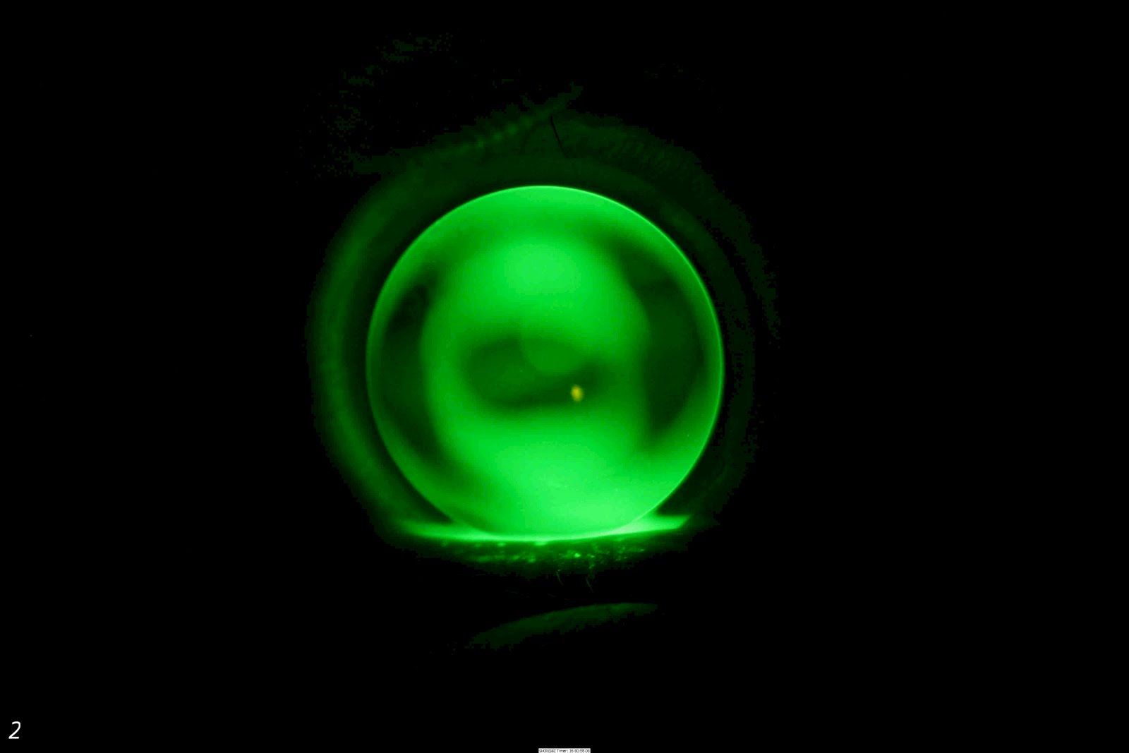
Sometimes this myelination continues into the eye, and is visible on the retina as bright white flame-shaped streaks which follow the course of the retinal nerve fibres and are situated superficially and often obscure the retinal blood vessels.

Do they affect vision? YES - they may cause field defects corresponding to their area of projection into the visual field. Should this person be driving? ...depends on their contralatteral eye!
This image demonstrates the path of the nerve fibre layer of the retina really well. 

Epiretinal Membrane and Pseudohole: Caught in the Act

Occasionally, an epiretinal membrane (a thin sheet of fibrous tissue) will form on the inner limiting membrane interface on the retina, contract over time and form what looks for all the world like a macular hole. These require no treatment unless acuity is poor enough- then in the skilled hands of a vitreoretinal surgeon the epiretinal membrane can be peeled off. 

Here are some great images of this membrane and the resultant pseudohole which we were able to image in the practice recently. It required no attention at present, but we'll watch it regularly. 


The Zeiss Cirrus HD-OCT allows us to cut sections through the imaged tissue to explore the retina in 3-D.

Debug these RGP Fits

These are some poorly fitting RGP contact lenses, taken during the fitting process. See if you can work out what needs to be adjusted to improve the fitting.