Highlights

Wednesday, March 30, 2011

Cellphone Photodocumentation

About a year ago I started doing a day a week at a satellite practice. While it is pretty well equipped I soon missed the imaging suite we have at Black Gates in Lower Hutt.
I now routinely photodocument as much as I can. This practice is far superior to sketching, or a brief description.
The reality is that we all have a reasonable microcamera within arms reach most of the time, and with a little practice the optics can be aligned to take images through the slitlamp optics.
In the last months I have used this to document the following:
- lid lesions
- conjunctival inflammations and lesions
- RGP fits
- contact lens deposits
- corneal scars and infiltrates
- iris naevii and abnormalities
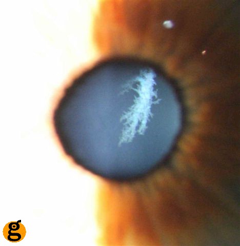
- lens opacities (cortical are easiest)
- optic cupping and Drance hemorrhages
- choroidal naevii
- choriotinal scars
- Weiss rings and floaters

Attaching the images to the patient record is easy- just email them to be practice, cut and paste and there you go.

Have fun

Greg


Wednesday, September 1, 2010

Cataract Zoo 5: Anterior Polar Cataract (with Iris Strands)

This is an interesting one!

This is a contact lens patient of mine - an incidental finding. She is moderately myopic (-7.00) and does well in SiHy lenses. She also has some WWP (white without pressure) in her retinal mid-periphery, which looks like a flying lizard. Cool!


Iris Strands linking the iris collarrette to the site of the Anterior Polar Cataract.


Dr Google helped me discover this in the text book "Pediatric Ophthalmology and Strabismus": (Kenneth Weston Wright; Peter H. Spiegel). They describe this as a small discrete opacity usually less than 3mm in diameter, usually in the centre and on the anterior capsule of the lens, not usually visually significant and not progressive.
It is caused by and abnormal separation of the lens vesicle from the surface ectoderm during embryonic lens development.
It is Bilateral in about 1/3 of cases; about 1/3 of patients will also have STRABISMUS, ANISOMETROPIA or AMBLYOPIA.
It is exhibits autosomal dominance in about 10% of cases.

Ddx: Peters anomaly - adheres to the cornea; abnormal angle; glaucoma risk.


Sunday, August 29, 2010

Cataract Zoo 4: Christmas Tree Cataract


A Christmas Tree Cataract is comprised of refractile needles located in the deep cortex of the lens. They crisscross the lens fibres reflecting light in different colours, looking very much like tinsel on a christmas tree..hence the name. 

The cause is thought it be from the age related breakdown of lens crystallins induced by elevated calcium levels. 

G. Adrien Shun-Shin* Gijs F. J. M. Vrensen,\ Nicholas P. Brown,* Ben Willekens,^
Marianne H. Smeets,^ and Anthony J. Bron*)

Cataract Zoo 3: Posterior Supcapsular

Posterior Subcapsular Cataract:
The greasy reflection off the back surface of the lens is the subcapsular opacity, and was visually devastating. This patient is diabetic and while this was the worst eye -there were similar changes in their other eye. This type of lens opacity is a common consequence of Diabetes. Modern surgical techniques gave this patient spectacular vision once again.

Cataract Zoo 2 : Dendritic Cataract

Dendritic Cataract:
This is a congenital cataract. An opacity in the sutures of the lens often has intricate branches. This one is beautifully tinged with azure. The vision is only one letter different in this eye. 

Cataract Zoo 1: Cortical

Cortical Changes:  Cataracts are ubiquitous - often the first obvious signs are cortical.

Cortical changes revealed by dilated pupil
Vesicles: fluid filled bubbles
More Bubbles: retro-illuminated
Vesicles with some sutural spokes (and a spec of posterior subcapsular change): 2 lines VA reduction


Myelinated Nerve Fibres

The insulating and protective fatty myelin sheath, which surrounds our nerves to protect and support them, doesn't generally enter the eye.
This myelination of the optic nerve develops embryologically in a posterior to anterior direction. The nerves are roughly myelinated in the intracranial and intracanal portions of the optic nerve by the seventh month (after conception) but doesn't usually reach the lamina cribosa until full term.
Sometimes this myelination continues into the eye, and is visible on the retina as bright white flame-shaped streaks which follow the course of the retinal nerve fibres and are situated superficially and often obscure the retinal blood vessels.

Do they affect vision? YES - they may cause field defects corresponding to their area of projection into the visual field. Should this person be driving? ...depends on their contralatteral eye!
This image demonstrates the path of the nerve fibre layer of the retina really well.